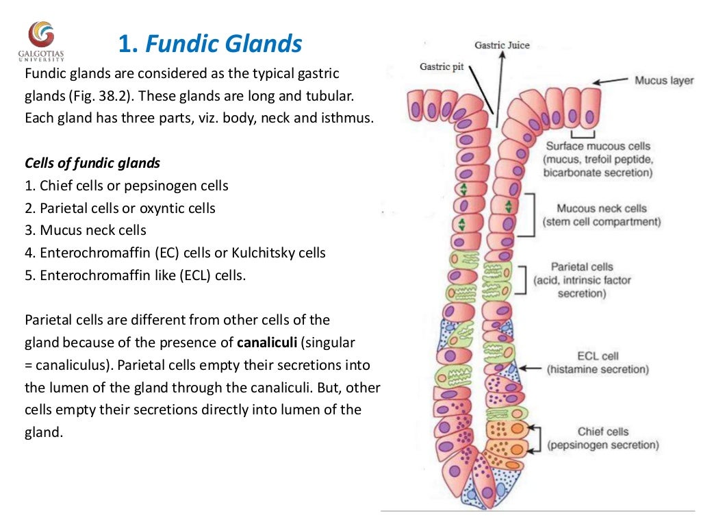
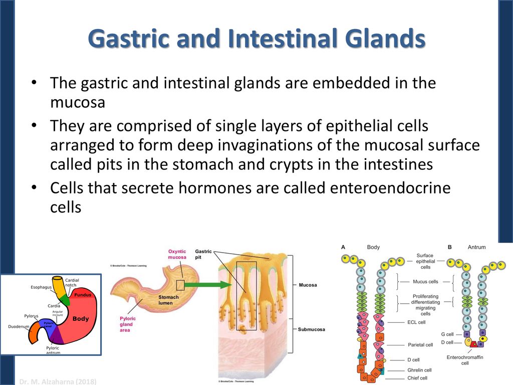
Stomach Gastric Glands Biology Diagrams The stomach lining consists of distinct regions with specialized functions, including the antral and oxyntic mucosa. These areas differ in structure, cellular composition, and physiological roles, contributing to digestion and maintaining gastric health. Gastrin-producing G cells reside in the deeper portions of the gastric glands and Gastric glands are glands in the lining of the stomach that play an essential role in the process of digestion.Their secretions make up the digestive gastric juice.The gastric glands open into gastric pits in the mucosa.The gastric mucosa is covered in surface mucous cells that produce the mucus necessary to protect the stomach's epithelial lining from gastric acid secreted by parietal cells

Gastric gland and gastric wall: histology diagram. Gastric glands open into the base of gastric pits. They are found throughout the entire inner surface of the stomach and are divided into 3 types depending on the region in which they are found. Gastric glands proper (principal glands) are found in the fundus/body of the stomach. The cells of In the inner surface layer (epithelium) of the stomach, gastric glands produce mucus to protect the stomach lining, hydrochloric acid for digestion and digestive enzymes such as pepsinogen However, in people with gastric cancer, some of the glands from the normal, non-cancerous stomach lining showed changes under the microscope that resembled the early stages of transitioning to cancer.
Scientists uncover hints of a potential new cause of stomach cancer Biology Diagrams
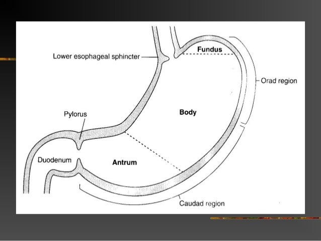
gastric gland, any of the branched tubules in the inner lining of the stomach that secrete gastric juice and protective mucus.. There are three types of gastric glands, distinguished from one another by location and type of secretion. The cardiac gastric glands are located at the very beginning of the stomach; the intermediate, or true, gastric glands in the central stomach areas; and the
Gastric juice is secreted by gastric mucosal glands, and contains hydrochloric acid, mucus, and proteolytic enzymes pepsin (which breaks down proteins), and lipase (which breaks down fats). When the stomach is empty, and not distended, the lining is thrown up into folds called rugae. After eating, these folds flatten, and the stomach is able to
Mucous Cells, Parietal Cells & Chief Cells - Britannica Biology Diagrams
Each element contributes to how effectively the stomach performs its duties. Gastric Mucosa Structure. The gastric mucosa, a specialized lining of the stomach, serves as a barrier and a site of active secretion. This mucosal layer is composed of distinct regions, each with unique cellular compositions and functions.
